Pelomyxa palustris
Pelomyxa palustris, almost 2 mm in length

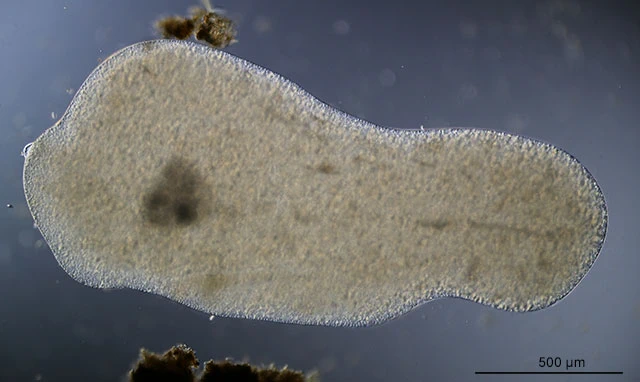
Pelomyxa palustris Greeff, 1874

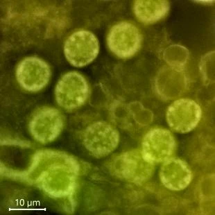
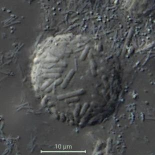
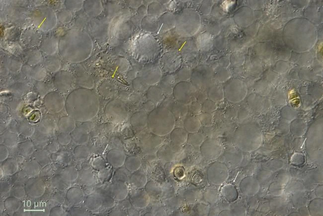
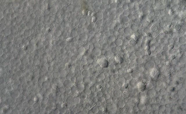
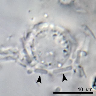
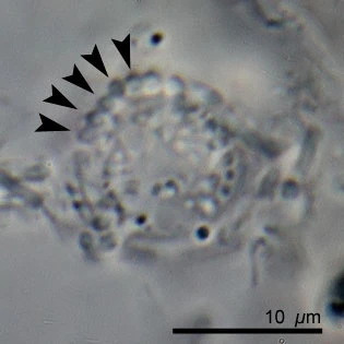
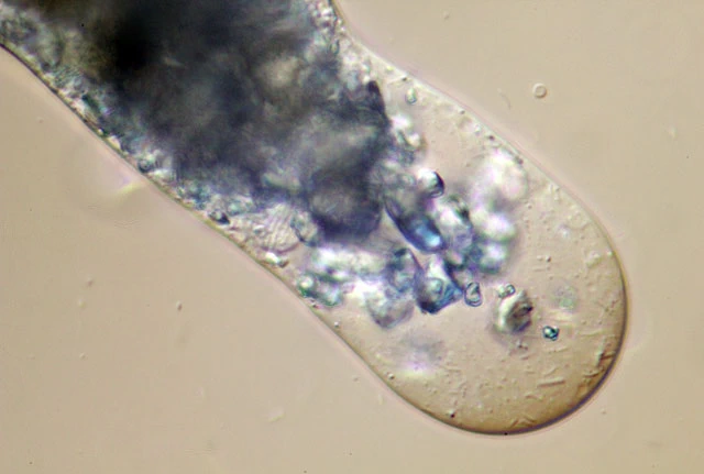
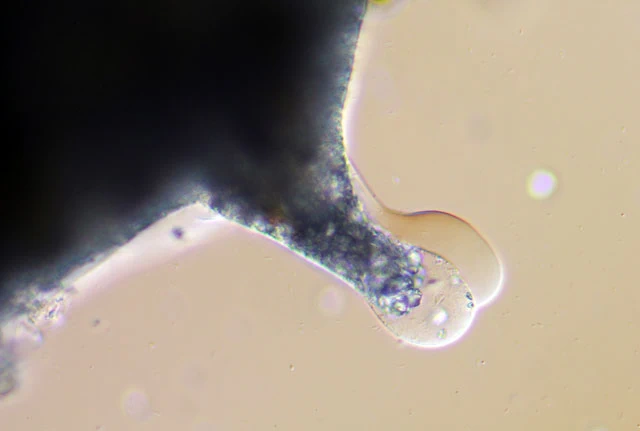
Diagnosis: Cells amoeboid, reaching 5 mm long with a large anterior pseudopod and a posterior uroid. With numerous nuclei. Short cilia have been demonstrated. Cytoplasm contains usually numerous glycogen bodies, as large or larger as the nuclei (10 µm). Nuclei surrounded by symbiotic bacteria. Symbiotic bacteria present.

Habitat: In sediments of swamped stagnant and low-flow freshwater reservoirs at a depth of 20 – 50 cm. Common. I also found numerous specimens in water filled tree hollows.

Remarks: A complex life cycle has been described. However, recent investigations (Frolov et al, 2004, 2005, 2006, 2011) indicate that this complex life cycle doesn’t exist and that the several stages in this life cycle are valid species, as is  Pelomyxa binucleata. The question is whether the name Pelomyxa palustris covers a distinct species or a morphotype-complex.

Pelomyxa palustris
Pelomyxa palustris
Nuclei (L) and a glycogen body (R) with adhering bacteria
Pelomyxa palustris
P. palustris, G = Glycogen bodies; N = Nuclei
Pelomyxa palustris
P. palustris, from a water filled tree hollow, Netherlands
Pelomyxa palustris
P. palustris, arrows indicate flagellae
Pelomyxa palustris
P. palustris, with flagellae
Pelomyxa palustris
P. palustris, g = glycogen body; n = nucleus
Pelomyxa palustris
P. palustris, nuclei (yellow arrows) and glycogen bodies (white arrows)
Pelomyxa palustris
Highly vacuolated cytoplasm with some nuclei and glycogen bodies
Pelomyxa palustris
Pelomyxa palustris
Nucleus, arrows indicate adhering bacteria and bacteria in cross section. Symbiotic bacteria
Pelomyxa palustris
P. palustris, 800 µm – Diepveen, Dwingeloo
Pelomyxa palustris
P. palustris, pseudopodium with bacteria
Pelomyxa palustris
Pelomyxa palustris, pseudopodium
Pelomyxa palustris
Pelomyxa palustris, about 2 mm in diameter
Ferry Siemensma, created March 1, 2019; last modified October 16, 2024
Recent posts